Artist in Residence - Diamond Light Source Blog
September 2008 - Festival of Science
The Festival of Science, held in Liverpool (European City of Culture this year) began for us on 9th September with a reception hosted by Diamond, Daresbury and STFC at the Bluecoat Gallery. Here are some of the final pieces of artwork were displayed.
These stainless steel sculptures made in collaboration with Paula Groves include textiles printed with iron filings in magnetic patterns. Finally they will be sited in the atrium in Diamond House.

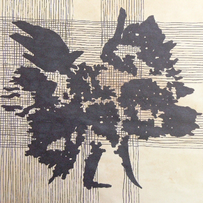
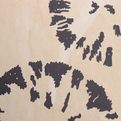
The wall panels below were inspired by the sail shapes of the Mary Rose were printed using iron pigments using pieces from the Diamond store. Backgrounds were stitched to echo the shadows created by the shapes. Frames made by Paula Groves.


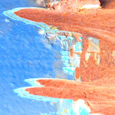
Right is a detail of a 2.5meter hanging, again inspired by the sail shapes of the Mary Rose but the fabric is dyed using rust and shows the difference in the depths of shade obtainable by using this mineral.
The following day Diamond's event, Worms Turn Detective! This was a series of three talks, the first, by Dr Mark Hodson of University of Reading explained how some earthworms can inhabit soil contaminated by metal and how they are assisting with the decontamination of this soil.
The second, by Dr Paul Schofield of Natural History Museum was looking at another solution to the same problem which was how phosphates, such as bonemeal, could immobilise the metal contamination.
Finally Dr Sam Shaw of University of Leeds presented on how toxic and radioactive contamination in the earth could be immobilised by the use of Green Rust. It is difficult to recreate condidions under which Green Rust is created as it requires low oxygen levels. However with the help of Diamond these conditions can be further investigated.
August 2008 - Work in Progress
Please click on a thumbnail for a larger image and further information: -


July 2008 - Natural History Museum
My second visit to the Natural History Museum to see Caroline Smith Curator of Meteorites. However this time she has prepared some iron minerals for me to photograph. Some of these are the crystals which are ground to give the powders used by Sam Shaw from University of Leeds in his experiments relating to decontamination of the earth. Also the pigments I hope to use in some of my final pieces.

Today I can look at the rocks under their microscope and take pictures of what I see. These inspirational images show the beautiful and varying structures of the different compounds.
I am particularly drawn to the cacoxenite image shown right both the vibrant gold colours and the starburst shapes of the crystals.
Cacoxenite is an iron phosphate found in iron rich soils and sediments.
The left hand image below is scorodite which contains iron and arsenic.
The central image below is of hermatite (one of several iron oxides) and hermatite in calcite.
 |
 |
 |
Many thanks to Caroline and the NHS, again, for all their help.
June 2008 - Mary Rose

It was a beautiful sunny day when we set off for the Portsmouth Historic Dockyard to see the Mary Rose, the historic artifacts that have been recovered from the vessel and visit the Historic Dockyard at Portsmouth.
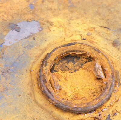
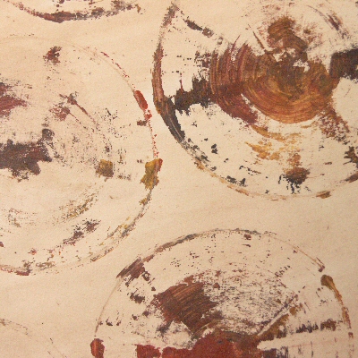
Scientists are not only working on the preservation of the timbers from the Mary Rose itself but also these artifacts and regularly visit Diamond to check on the status of their work. Since raising the vessel to the surface the oxidisation of the pyrites found in the fixings on the vessel and in many of the artifacts such as buckets or barrels, has caused acid which has adversly affected the timbers. In order to protect the wood from further deterioration, the team attempt to remove the pyrites before they turn to acid and the ship is continually sprayed to replace water.
Inspirational images taken at the Dockyard. Many thanks to Mark Jones and his team for their hospitality and assistance.
 |
 |
 |
May 2008
Have been having many thoughts about what I can do to tie the sciences from each of the beamlines up and provide a link between them and have finally come up with "The Project".
This will be to look at iron in some of its forms. Iron after all makes up about 5% of the earth's crust. Not only that, all the experiments I have looked at have involved iron in some form. From the largest meteorites to the tiniest particles in your brain, iron appears on the parchments of antiquity and in the timbers of the Mary Rose, in fact it is everywhere!
April 2008 - Parkinson's Disease
Following my meeting with Caroline I am now specifically looking at where iron is found in the body and any research is being done in this area. I go back to Liz Carpenter on the Protein Crysatlography beamline to see if there are any users looking at iron in the body that I could talk to. Unfortunately there aren't at the moment and I come away wondering how I can find my link.

However, one thing I have found during my time at Diamond is that you cannot plan specific meetings with beamline scientists and users to look at science you are interested in. While they are working on the beamlines time is precious and very often work is confidential so I am doubly grateful whenever anyone can spare time to talk to me so.
I am so lucky then when I am invited to meet with Joanna Collingwood from Keele University and Mark Davidson University of Florida who are comparing thin slices from a specific region in the brain from a group of 50% are healthy humans and 50% with Parkinson's disease (see image left). Not only that, but the final piece in my jigsaw, they are looking at iron concentrations!
The brain region is called the substantia nigra, and contains an important group of brain cells called dopaminergic neurones. These neurones are important for many functions, including enabling us to make controlled voluntary movements (i.e. they are "motor neurones").
A very exciting moment when they tell me they are looking at both the concentrations of iron and the different forms of iron (primarily different iron oxide structures) that are present in this region. It is already known that in Parkinson's sufferers there are fewer dopaminergic neurones in the brain as they are destroyed, but there are higher concentrations of iron in this region, and within the remaining dopaminergic neurones. This research is looking at where exactly this extra iron is found (also a range of other metals including zinc, calcium, copper, and manganese).
Is it more concentrated in the living cells or dying cells, or in the support cells? How much the iron in these cells is contained in ferritin (primary iron storage protein, which is only a few nanometres in diameter), and how much is found in neuromelanin, which is a pigment that can bind iron, and that is found in the cell bodies of the dopaminergic neurones. Also when the iron is found, what type of iron compound is it.
April 2008 - Meteorites

Meeting with Paul Schofield who is working on a project investigating the decontamination of soil affected by metals from mining and industrial sites. Today he is looking at a fragment of meteorite as it has many structural similarieties with the rocks here on earth.
The image on the right is a fragment from the Santa Catherina iron meteorite which fell to earth in Brazil between 10,000 and 40,000 years ago.
A meteorite is a fragment which has broken off an asteroid, a body which was formed at the same time as earth, also having a rocky surface and an iron nickel core with a molten layer around it. An iron meteorite is a piece from the core of the asteriod and can give insights into the secrets of our own planet's core. A piece from the rocky surface would be known as a stony meteorite.
As the molten iron nickel in the asteroid cools and solidifies different patterns of the various metal compounds are formed, each pattern unique to the temperature, pressure, rate of cooling and the oxygen present at the time. In time, rust (iron oxide) is formed on the surface of the meteorite, this too can give an indication of the various compounds that exist underneath the rust. The patterns formed by these compounds during this cooling process are known as phases.


Following this discussion, I feel I would like to learn more about the work being done in this area. So I arrange to meet Caroline Smith who is the curator of meteorites at the Natural History Museum. These images are of some of the stoney and iron meteorites she showed me. The green material in the meteorite shown left is gem quality olivine otherwise known as peridot a semiprecious gem used extensively in jewellery.
The small white mineral grains that can be seen in the stony meteorite below left contain calcium and aluminium. They have been used to date our solar system.
I am fascinated by the age and the distances these meteorites have travelled. The cinema has portrayed them as huge pieces of rock that are potentially going to destroy the earth. How does this compare with the tiny crystals I have seen here in different formations and the iron compounds that I know are inside our bodies?
 |
 |
 |
Many thanks to Caroline Smith and the NHS for their help. For more information, visit Natural History Museum
March 2008
More amazing conversations with scientists about materials they investigating on the beamlines. From meteorites to pigments every day is fascinating.
Once again though, I look at the scribbled notes and diagrams both my own and those the scientists have made to try and make clear to me these complicated processes. What happens to these fleeting moments when I not only comprehend the answer, but the question that I am asking so clumsily.
The only way this project makes sense is if there is some communication of these moments. Time is flashing past and I only have insights into tiny parts of completely disparate areas of research which are only linked by this fantastic machine. I don't want to be tied to one area and am searching for connections!
If only I can grasp the right questions, and keep on a single path then just maybe I will find a dialog that will stay with me and is worth communicating to a wider audience!
February 2008 - Protein Crystalography

Liz works for Imperial College London and has a lab here at Diamond where she assists the beamline scientists as well as customers of the College who are looking at Protein Crystalography, particularly proteins which sit on cell membranes.
Proteins are built from information held in DNA. This information is in the form of a key giving the order chemicals need to be assembled in. The beginning of the chain of chemicals will begin with an N and end with a C, hanging off each of these chemical blocks is an ? with another set of "hieroglyphic" symbols which look like the aerials on the heads of the teletubbies!
In diseases such as cancer some part of the code is incorrect, this will give a genetic disposition to the disease, but it may not manifest itself until some other portion of the dna becomes corrupted. Once this chain is assembled it folds into a recognisable structure (protein fold).
Proteins exist both inside and outside cells, these are relatively easy to decode as they are soluble in water.
The proteins that sit on the cell membrane are much harder to decode as they exist as part of the oily membrane. Very often their job is to push fluids such as sugars and other proteins, in and out of the cells. In viruses such as flu the membrane proteins are able to attach themselves to the surface of healthy cells and transmit materials that will cause the cell to reproduce the virus. Drugs such as Relenza are in fact other proteins which attach themselves to the membrane proteins of the flu virus making them unable to attach.
In order to try and grow crystals from these proteins, it is necessary to suspend them in a variety of materials such as alcohol, salt or detergents. The correct solution is all very much trial and error as in many cases these materials will stop the correct alignment of the proteins or in fact will form crystals themselves.
In her lab, Liz showed me how trays containing 96 different solutions which are filled by robot (see above left) with the same protein to see how each will grow. These crystals can take up to a year to form and will often be destroyed by the beam.
February 2008 - School of Optometry Cardiff
Another month has passed and what have I done ...
I have had a trip to a very modern minimal building in Cardiff, part of the University which houses the School of Optometry and Vision Services. Here (as well as part time film set for Dr. Who and Torchwood) PhD students and Professors study as well as performing research into eye diseases such as Keratoconis.

This research requires substantial amounts of funding, some of which is raised by the Eye Clinic, located on site and open the general public. In addition to performing eye examinations and dispensing, there are highly specialised clinics in low vision and assessing the needs of those who cannot communicate in the usual way. For more information regarding the Cardiff Eye Clinic Cardiff Eye Clinic
On a more scientific note, it was amazing to see their electron microscope, which sits on a bench in one of the labs! This is where the initial scientific investigation begins before progressing to the room sized beamlines at Diamond! The data from the Keratoconis experiments performed at Diamond in December has not yet been processed but I wonder how those images would compare to ones produced on the equipment here. Is this even possible and if so would they relate in any visible way to each other?
January 2008
After what seems like an age for Christmas, I am back! Have spent more time this month with the scientists discussing how they contribute to the analysis of the data, both in the setup of samples and beamlines and in processing the data to produce as accurate information as possible for the users. I have even been taken back to schoolgirl maths and relearnt integration calculus! Sheets of scribbled explanations, graphs and diagrams are produced to help me understand. For me, these are precious works of art in themselves, without the fluency of these delicate marks I would have no comprehension of this complicated new language.

I also saw a variety of the detectors that are used on the beamlines and watched how they are setup and calibrated.
The Germanium detector, checks the strength of the xray and the intensity of the patterns. It is used on the spectroscopy beamline. A small device which emits xrays is used to check the machine. The CCD detector works like a camera and creates organised patterns of the xrays, and the scintillator detectors shown here can be installed singly or as an array, and is used to count the dots on each pulse of light.
Other detectors which are built in these labs are so minute they can only be successfully assembled under a magnifying lamp, where the gold tape which carries the signal from the detector to the amplifier is positioned with patience and delicacy.
I feel at home in these labs, I can relate to the materials and tools used here and wonder if they could be used in the final pieces.

Coincidentally, there were some images of the detectors used in the Large Hadron Collidor at Cern in the newspaper this week, these are much larger than those at Diamond, but the component parts form fascinating symmetries and patterns. These inspire me further and next month I am hoping to be able to look inside the detectors and to use some of these same materials in my own work!
December 2007 - Nanoscience

Today I have a meeting with Sharnjeet Dhesi Principle Scientist on the Nanoscience beamline I06. Here users are looking at surfaces rather than structures at atomic level 10-9 known as a nanometer.
On this beamline it is possible to get images of the surface patterns or videos immediately rather than processing diffraction patterns through software.
Surfaces are never completely flat but made up of atomic steps where each step is made up from layers of atoms. One strand of investigation carried out on this beamline is looking at the reaction of gases on these surfaces (typically oxygen on iron easy which is relatively easy to understand). Metals are heated until they are gases and then condensed onto surfaces such as silicon.
Another, is a set of experiments on semiconductors. Observations are then applied to superconductors which can only work at very low temperatures and are therefore harder to set up. If successful could replace silicon chips with another element such as germanium.
December 2007 - Keratoconus

The first experiments we see are an investigation for Treatments of Keratoconus on I22 beamline Calibration was underway using rat collagen in preparation for Cardiff University who are looking at the treatment of keratoconus. This is a disease affecting the collagen in the cornea. The cornea is responsible for transmiting and focusing the light coming into the eye and the symptoms of this disease include glare, corneal astigmatism and increased sensitivity to light. How simple the setup of this experiment appears compared to the complexity and sophistication of the synchrotron itself! This image shows sections of cornea wrapped protected by cheap clingfilm, apparently the most suitable material! Readings are taken in an 11 x 11 grid across each of the samples.
The cornea is made up of about 200 layers of collagen fibres, each layer has fibres lying in a different direction to give strength and rigidity, in sufferers of keratoconus these layers have slipped out of place. Cardiff are investigating the use of riboflavin (vitamin B2) which interacts with the UV light so rather than damaging the fibres it forms a glue and provide crosslinks between the fibres.
Immediately I can relate to the shared properties of a piece of textile and the fibres in a cornea and how the concept could be visualised. For more information visit Cardiff University Optometry
Well, the end of the first month, I can now get to the studio without doing an extra circuit of the ring and have learnt more about science in 4 days at Diamond than I did in 5 years at school!
I have talked to scientists with a huge diversity of expertise. My initial impression is of how isolated each of the experiments is. The synchrotron is a circular structure, but each beamline fractures the ring. As more beamlines come on board, the more broken the structure is likely to become. An important part of the project, for me, is to provide a thread showing how all the aspects of this science fit together as part of a bigger picture.
December 2007 - Beginnings
I can’t believe its time to begin the residency. We meet with some of the scientists who are prepared to explain some of the work they are doing and what it will mean to us on a day to day basis.

There is so much happening here and I have absolutely no idea where to begin. I don’t even know the questions that I need to ask! I turn up for my first day’s proper work, and feeling apprehensive I use my shiny new pass to obtain access to the synchrotron and our studio. Immediately, in the giant ring for the first time on my own, I feel disorientated, but confidently stride two circuits, hoping noone will notice before I locate the studio and safety.
We have a good sized room but it feels impersonal and characterless. The sooner we get some work on the walls the sooner I will feel at home and that we have made a start.